

Eye Disease
Red eye, visual disturbance, glaucoma, cataracts and ophthalmology emergencies.
Last reviewed
Use this quick reference guide to study and ensure you have mastered these common cases from the curriculum. You can generate specific cases on our SCA cases page or even practice them with our specialised and UK guidelines trained AI Actors.

Table of Contents
1Red Eye: Differential Diagnosis and Management
Differentiating between benign and sight-threatening causes is crucial.
Visual explanation
Painful red eye: ciliary flush versus corneal infiltrate
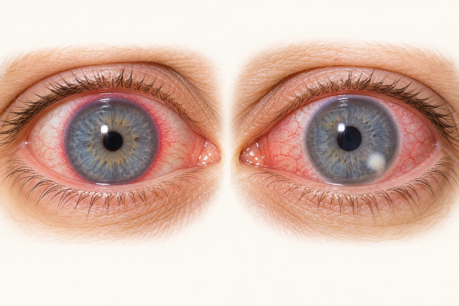
- Ciliary flush around the limbus
- Focal white corneal infiltrate
Anterior uveitis example
Pain, photophobia and blurred vision support suspicion. Ciliary flush is not specific to uveitis; specialist examination is needed.
Microbial keratitis example
Painful red eye with a corneal opacity, especially in a contact lens wearer: arrange same-day emergency ophthalmic assessment.
Remember: Check visual acuity in each eye, contact lens use, pupils and photophobia. Suspected uveitis also needs urgent ophthalmic assessment. Do not start steroid eye drops empirically in an undiagnosed red eye.
AI-generated teaching illustrations, not patient photographs. Findings may be subtle or absent in practice. Sources: College of Optometrists: anterior uveitis · College of Optometrists: microbial keratitis · NHS: uveitis · NHS formulary: steroid eye drops
Conjunctivitis
- Bacterial: Purulent discharge, eyes stuck together in the morning. Treat with topical chloramphenicol if severe or not resolving (though often self-limiting).
- Viral: Watery discharge, often associated with URTI, pre-auricular lymphadenopathy. Highly contagious. Conservative management.
- Allergic: Itch is the predominant symptom, bilateral, watery discharge. Treat with topical antihistamines/mast cell stabilisers.
Subconjunctival Haemorrhage
Bright red blood under conjunctiva, painless, normal vision. Reassure (check BP/INR if recurrent or on anticoagulants).
Red Flags (Refer Urgent/Same Day)
- Pain: Moderate to severe pain (suggests scleritis, keratitis, uveitis, or acute glaucoma).
- Photophobia: Suggests iritis/uveitis or keratitis.
- Reduced Visual Acuity: Suggests corneal or intraocular involvement.
- Contact Lens Wearers: High risk of microbial keratitis (pseudomonas). Refer same day.

2Sudden Loss of Vision: Assessment and Red Flags
Sudden visual loss (monocular or binocular) is an emergency until proven otherwise.
Visual explanation
Acute visual loss: CRAO versus CRVO
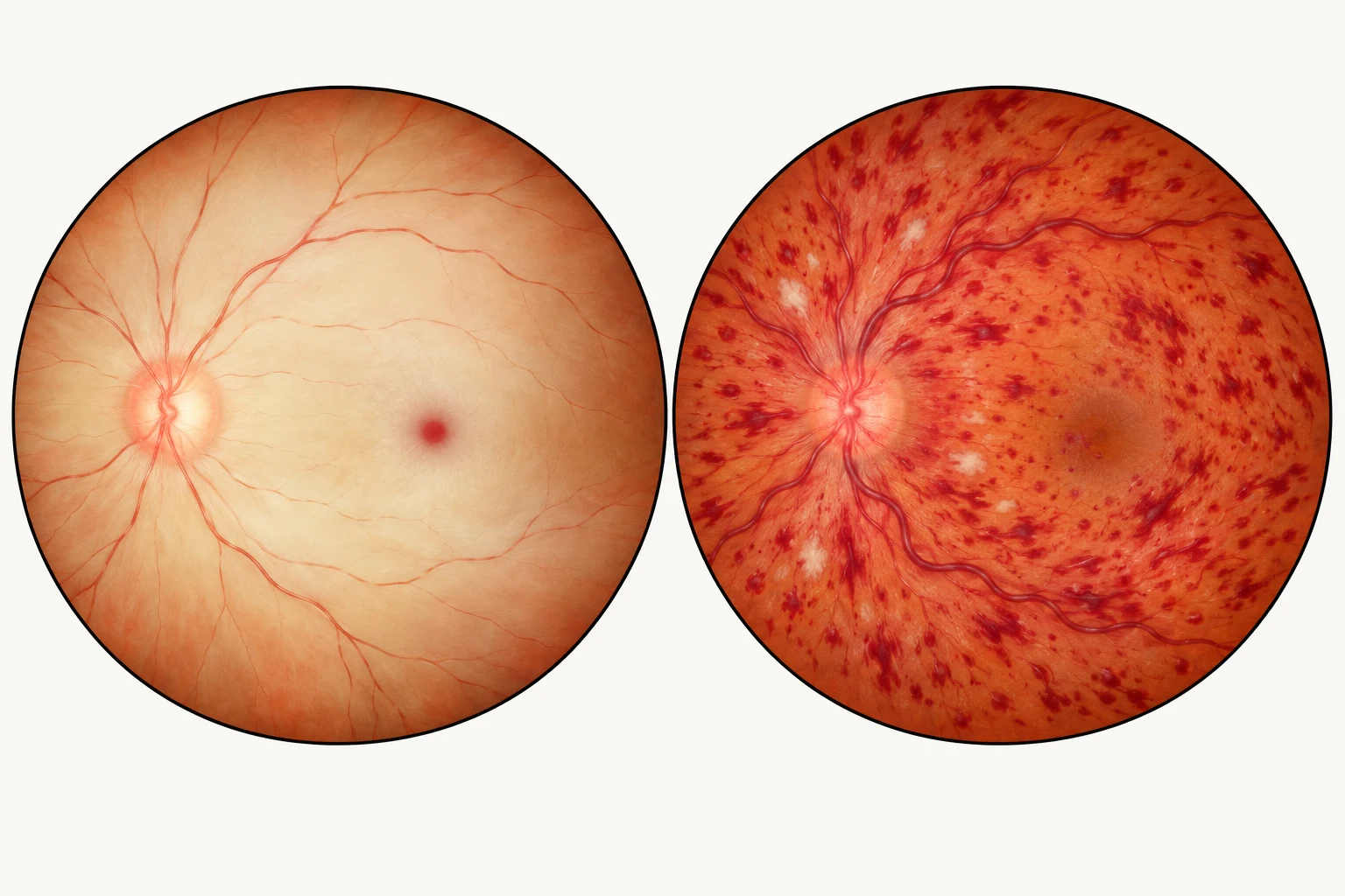
- Retinal pallor / ischaemic whitening
- Cherry-red foveal spot
- Widespread retinal haemorrhages
- Dilated, tortuous retinal veins
Central retinal artery occlusion
Sudden, often profound monocular loss. Activate the emergency ophthalmic / stroke pathway immediately; a normal early fundus does not exclude it.
Central retinal vein occlusion
Haemorrhages in all four quadrants with venous dilatation and tortuosity. Arrange urgent ophthalmic assessment and review vascular risk factors.
Remember: Acute visual loss needs immediate assessment. Do not delay referral to obtain a classic fundus view. Ask about GCA symptoms in people over 50, and act urgently if suspected.
AI-generated schematic fundus views, not patient photographs. Classic arterial changes may take hours to appear. Sources: AAO: retinal artery occlusion · AAO EyeWiki: early fundus findings · RCOphth: retinal vein occlusion guideline
Painless
- Amaurosis Fugax: Transient monocular blindness ("curtain coming down"). TIA management (Aspirin 300mg, refer to TIA clinic).
- Central Retinal Artery Occlusion (CRAO): Pale retina with "cherry red spot". Sudden, profound loss of vision. Ophthalmic emergency.
- Central Retinal Vein Occlusion (CRVO): "Stormy sunset" appearance (haemorrhages).
- Retinal Detachment: Flashes (photopsia), floaters, and field loss ("shadow/curtain"). Urgent referral (same day/24h).
- Vitreous Haemorrhage: Sudden loss of vision, often in diabetics.
Painful
- Optic Neuritis: Pain on eye movement, reduced colour vision (red desaturation), central scotoma. Associated with MS.
- Acute Angle Closure Glaucoma: (See Section 6).
- Giant Cell Arteritis: (See Section 7).
3Gradual Loss of Vision: Cataracts, AMD, and Glaucoma
The "big three" causes of gradual visual decline in the elderly.
Assessment
Check Visual Acuity (VA) with distance glasses and pinhole (to correct refractive error). Check Red Reflex.
Cataracts
Opacity of the lens.
- Symptoms: Glare (dazzle from headlights), blurred vision, "washed out" colours.
- Management: Refer for surgery when symptoms affect lifestyle/driving.
Refractive Error
Most common cause. Improves with pinhole. Refer to Optometrist.
4Age-Related Macular Degeneration (AMD)
The leading cause of blindness in the UK. Affects central vision.
Dry AMD
Slow, gradual decline.
- Signs: Drusen (yellow deposits) on the macula.
- Management: No cure. Lifestyle advice (stop smoking, diet/supplements). Register as visually impaired if eligible.
Wet AMD
Rapid deterioration, distortion of straight lines (metamorphopsia).
- Signs: Haemorrhage or fluid at the macula.
- Action: Urgent referral (2-week pathway) to a macular clinic.
- Treatment: Anti-VEGF intravitreal injections can save sight.
5Chronic Open Angle Glaucoma (COAG)
Asymptomatic in early stages (insidious loss of peripheral vision).
Risk Factors
Age, Family History (first-degree relative), Black ethnicity, Myopia.
Diagnosis
Triad of: 1. Raised Intraocular Pressure (IOP), 2. Visual Field Defect, 3. Optic Disc Cupping.
Screening
Rely on opticians. Over 40s should have checks every 2 years.
Management
- Prostaglandin Analogues (e.g., Latanoprost): First-line (increase outflow). Side effects: longer eyelashes, iris pigmentation.
- Beta-blockers (e.g., Timolol): Reduce production. Caution in asthma/heart block.
Note: DVLA must be notified.
6Acute Angle Closure Glaucoma
Ophthalmic emergency. Closure of the drainage angle causing rapid IOP rise.
Symptoms
Severe ocular pain, headache, nausea/vomiting, blurred vision, haloes around lights.
Signs
Red eye, hazy (oedematous) cornea, fixed mid-dilated pupil, eye feels "rock hard".
Management
- Immediate: 999/Same-day admission to Ophthalmology.
- Primary Care: If significant delay expected, start Pilocarpine drops (constricts pupil), Acetazolamide 500mg IV/PO (if available/advised), Analgesia, Antiemetics.
7Giant Cell Arteritis (Temporal Arteritis)
Vasculitis affecting large vessels. Major risk is permanent blindness due to ischaemic optic neuropathy.
Red Flags
New headache >50 years, scalp tenderness (pain when brushing hair), jaw claudication (pain on chewing), visual disturbance (diplopia/blurring).
Investigation
ESR/CRP (usually markedly raised).
Suspected GCA - NO Visual Symptoms
Urgent referral (e.g., Rheumatology/GCA clinic). Start Prednisolone 40-60mg daily.
Suspected GCA - WITH Visual Symptoms
Same-day emergency ophthalmology referral. Start high-dose steroids (e.g., IV Methylprednisolone or oral 60-100mg) immediately. Do not wait for biopsy.

8Blepharitis and Dry Eye Syndrome
Very common chronic conditions causing discomfort.
Blepharitis
Inflammation of eyelid margins (Staph aureus or Seborrhoeic).
- Symptoms: Gritty, burning, crusting on lashes (morning).
- Management: Lid Hygiene (warm compresses, massage, cleaning with baby shampoo/bicarb) twice daily. Topical antibiotics (Chloramphenicol ointment) if signs of acute infection (severe inflammation).
Dry Eye Syndrome
Tear deficiency or evaporative loss.
- Symptoms: Gritty, "sand in eyes", reflex tearing.
- Management: Artificial tears (Hypromellose, Carbomers). Ointments at night. Preservative-free drops if used frequently (>4 times/day).
9Eyelid Lumps: Chalazion vs. Stye
Differentiating common lid swellings.
Stye (Hordeolum)
Acute infection (abscess) of a lash follicle (external) or Meibomian gland (internal).
- Features: Painful, red, tender lump at lid margin.
- Treatment: Warm compresses, epilation of lash. Topical antibiotics only if associated conjunctivitis/cellulitis.
Chalazion (Meibomian Cyst)
Chronic sterile granulomatous inflammation of Meibomian gland.
- Features: Firm, painless (or mildly tender) round lump within the lid (not at the margin).
- Treatment: Warm compresses and massage (most resolve in months). Referral for incision and curettage if persistent and bothersome (check local funding criteria).
10Diabetic Eye Disease: Screening and Retinopathy
Diabetic retinopathy is a microvascular complication causing blindness.
Screening
All diabetics >12 years offered annual photographic screening (DES).
Stages
- Background (R1): Microaneurysms, dot/blot haemorrhages. Observation.
- Pre-proliferative (R2): Cotton wool spots (ischaemia), venous beading. Closer monitoring.
- Proliferative (R3): New vessels (neovascularisation). High risk of bleed/detachment. Urgent laser photocoagulation needed.
- Maculopathy: Exudates/oedema near the fovea. Threatens central vision.
GP Role
Optimise HbA1c (target usually 48-53 mmol/mol) and BP (<140/80 or <130/80 if end-organ damage) to slow progression.
Practice with AI Actors
3 relevant roleplay cases
Apply what you've learned about Eye Disease in a realistic, voice-driven consultation. Speak with the AI Actor as you would a real patient and get instant feedback.

Related Topics
ENT
Ear, nose and throat conditions including hearing loss, sore throat, sinusitis and vertigo.
Women's Health
Contraception, menstrual problems, menopause, pregnancy-related issues and gynaecological concerns.
Sexual Health
STI management, sexual dysfunction, LGBTQ+ health and contraception counselling.