

ENT
Ear, nose and throat conditions including hearing loss, sore throat, sinusitis and vertigo.
Last reviewed
Use this quick reference guide to study and ensure you have mastered these common cases from the curriculum. You can generate specific cases on our SCA cases page or even practice them with our specialised and UK guidelines trained AI Actors.
Table of Contents
1Sudden Sensorineural Hearing Loss (SSNHL)
This is an otological emergency. Patients often describe a "blocked ear" (like cotton wool) or a sudden drop in hearing (often upon waking).
Visual explanation
Sudden hearing loss: the referral clock
Adult hearing loss not explained by an external or middle-ear cause
Onset within 3 days · in past 30 days
Refer immediately to ENT or an emergency department.
↳ Assessment within 24 hours
Sudden onset · more than 30 days ago
Refer urgently to ENT or audiovestibular medicine.
↳ Assessment within 2 weeks
Rapid worsening · over 4–90 days
Refer urgently to ENT or audiovestibular medicine.
↳ Assessment within 2 weeks
Remember: A blocked-ear sensation can be sensorineural loss. Document onset precisely and examine the ear. Do not postpone referral for routine audiometry. Associated focal neurological signs may require a stroke pathway.
Simplified teaching diagram. Sources: NICE NG98: sudden hearing loss
Assessment
Differentiate from conductive loss (wax/effusion) using tuning forks:
- Weber's Test: Localises to the good ear in Sensorineural loss (localises to the bad ear in Conductive loss).
- Rinne's Test: Air conduction > Bone conduction (Normal/Positive) in Sensorineural loss.
Action
- Referral: Immediate referral to ENT (to be seen within 24 hours/same day).
- Treatment: High-dose oral steroids (e.g., Prednisolone 60mg) initiated early (ideally <72 hours) significantly improve prognosis.
Do not treat as "Eustachian Tube Dysfunction" without excluding this.

2Otitis Media and Glue Ear
Common in children. Most cases are viral and self-limiting.
Visual explanation
Otoscopy: bulging drum versus middle-ear effusion
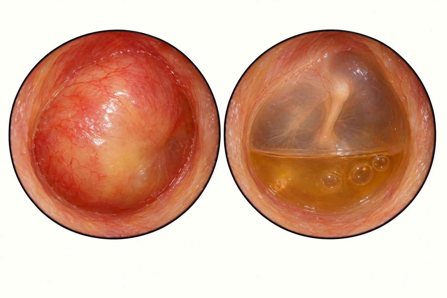
- Opaque, bulging tympanic membrane
- Air–fluid meniscus behind the drum
- Air bubbles within the effusion
Acute otitis media example
A bulging, opaque drum supports AOM in an acute illness. Redness alone is insufficient.
Otitis media with effusion example
Amber fluid, an air–fluid level or bubbles can explain conductive hearing loss without an acute infection.
Remember: Interpret the drum alongside pain, fever, hearing and systemic condition. Appearance alone does not determine antibiotic need. Persistent hearing difficulty needs an appropriate hearing assessment.
AI-generated teaching illustrations, not patient photographs. Appearances vary; use the clinical context. Sources: NHS: glue ear · NICE NG91: acute otitis media
Acute Otitis Media (AOM)
Antibiotics: Usually not indicated. Prescribe immediate antibiotics if:
- • Systemically unwell.
- • High risk of complications (immunocompromise, CF).
- • Otorrhoea (discharge from perforation).
- • Under 2 years old with bilateral infection.
Delayed Script: Consider for those who do not improve after 3 days.
Otitis Media with Effusion (Glue Ear)
- Presentation: Hearing loss, behavioural changes, speech delay. Dull, retracted drum with fluid level/bubbles.
- Management: Active observation for 3 months (most resolve). Refer for hearing assessment/grommets if persistent >3 months and hearing loss is significant.
3Otitis Externa and "Malignant" Otitis Externa
Inflammation of the ear canal ("Swimmer's Ear").
Simple Otitis Externa
Pain, itch, discharge. Canal is swollen/red.
- Treatment: Aural toilet (microsuction) if blocked. Topical antibiotic + steroid drops (e.g., Gentisone/Sofradex) for 7-14 days. Keep ear dry.
- Swabs: Take if treatment fails.
Malignant (Necrotising) Otitis Externa
Life-threatening osteomyelitis of the skull base (usually Pseudomonas).
- Red Flags: Elderly, Diabetic or Immunocompromised patient with severe, unremitting pain (worse at night) and granulation tissue in the canal floor.
- Action: Urgent ENT referral/Admission. Requires systemic antibiotics (Ciprofloxacin) and imaging.
4Tinnitus: Assessment and Red Flags
Perception of sound in the absence of external stimuli.
Assessment
Is it unilateral or bilateral? Is it pulsatile?
Red Flags (Refer to ENT)
- Unilateral Tinnitus: Suspect Acoustic Neuroma (Vestibular Schwannoma). Requires MRI.
- Pulsatile Tinnitus: Suspect vascular cause (e.g., Glomus tumour, Carotid stenosis).
- Associated Unilateral Hearing Loss.
Bilateral Tinnitus
Usually age-related or noise-induced. Check BP and medication review (aspirin/NSAIDs/loop diuretics). Management is supportive (masking devices, CBT).
5Vertigo: Differentiating BPPV, Labyrinthitis, and Meniere's
True vertigo is an illusion of movement (spinning).
Visual explanation
Dix–Hallpike and Epley: a right-sided positioning sequence
Right posterior-canal example. Steps 1–2 show the Dix–Hallpike position. Continue through 3–5 for Epley only after confirming the diagnosis, side and suitability.
Swipe through the five positions →

1. Turn toward the tested ear
Sit upright. Turn the head 45° to the patient’s RIGHT.
2. Lie back and observe
Keep the right turn. Support the head in about 20° of neck extension; observe the eyes.
3. Turn the head LEFT
From position 2, turn the head 90° left without lifting it. Keep the body supine.
4. Roll onto the LEFT side
Roll the body and head left; direct the nose diagonally down, about 45° toward the bed.
5. Sit up on the LEFT
Guide the patient up from the left side with the chin gently tucked.
Dix–Hallpike interpretation: look for characteristic transient upbeating, torsional nystagmus, with the upper poles beating toward the tested ear. Dizziness alone does not establish posterior-canal BPPV.
Epley timing: hold each lying position for at least 30 seconds and until provoked vertigo settles; follow the local protocol.
AI-generated positioning illustrations; angles are approximate. RIGHT and LEFT always refer to the patient.
Open the full positioning sheetRemember: Use only if trained and positioning is safe. Do not perform Epley with unstable cervical spine disease; use an appropriate referral pathway. Reverse directions only for confirmed left-sided posterior-canal BPPV.
Simplified teaching diagram. Sources: NICE NG127: positional vertigo · AAO-HNS: BPPV diagnosis · RCGP: repositioning technique · NHS: right-sided Epley
Benign Paroxysmal Positional Vertigo (BPPV)
- Features: Vertigo triggered by head movement (rolling in bed, looking up). Lasts seconds. No hearing loss.
- Diagnosis: Positive Dix-Hallpike manoeuvre (rotatory nystagmus).
- Treatment: Epley manoeuvre.
Labyrinthitis / Vestibular Neuritis
- Features: Acute, severe continuous vertigo (days/weeks). Often follows viral illness. Labyrinthitis includes hearing loss; Neuritis does not.
- Treatment: Prochlorperazine (short course only, max 3 days) to avoid inhibiting central compensation.
Meniere's Disease
- Triad: Vertigo (hours) + Tinnitus + Sensorineural Hearing Loss (fluctuating) + Aural fullness.
- Management: Betahistine prophylaxis. Referral to ENT.
6Acute Rhinosinusitis and Nasal Polyps
Inflammation of the nose and paranasal sinuses.
Acute Sinusitis
Usually viral.
- Bacterial: Suspect if symptoms >10 days, "double sickening" (worse after initial improvement), purulent discharge, severe local pain, fever >38°C.
- Treatment: High-dose steroid nasal spray (Mometasone) for 14 days. Penicillin V or Doxycycline if severe/bacterial.
Nasal Polyps
- Red Flag: Unilateral polyp requires urgent referral (suspect malignancy).
- Bilateral: Treat with steroid drops/sprays. Refer if obstructive.
7Sore Throat: Centor/FeverPAIN and Quinsy
Differentiating viral from Group A Strep infection.
Centor / FeverPAIN Score
Score criteria (Fever, Purulence, Attend rapidly <3 days, Inflamed tonsils, No cough):
- Score 4-5: Consider immediate antibiotics (Phenoxymethylpenicillin 500mg QDS or 1g BD for 10 days).
- Score 2-3: Delayed prescription.
Quinsy (Peritonsillar Abscess)
- Features: Severe unilateral pain, trismus (difficulty opening mouth), "hot potato" voice, deviated uvula (pushed away from abscess).
- Action: Immediate admission for IV antibiotics and drainage.

8Hoarseness and Laryngeal Cancer
Any unexplained hoarseness requires investigation.
2-Week Wait Referral
Aged 45 and over with:
- • Persistent unexplained hoarseness (>3 weeks).
- • An unexplained lump in the neck.
Primary Care Investigation
Consider Chest X-ray (to exclude apical lung tumour causing recurrent laryngeal nerve palsy) while awaiting ENT referral.
9Dysphagia vs. Globus Pharyngeus
Difficulty swallowing vs. sensation of a lump.
Dysphagia (Difficulty Swallowing)
- Red Flags: Progressive (solids then liquids), weight loss, pain on swallowing (odynophagia).
- Action: Urgent referral (2-week wait) for upper GI endoscopy (to exclude oesophageal cancer).
Globus Pharyngeus
- Features: Sensation of a lump in the throat. Swallowing is normal (often relieved by swallowing food/drink). No pain. Intermittent.
- Management: Reassurance (diagnosis of exclusion). Treat reflux if present. Refer if persistent or diagnostic uncertainty.
10Head and Neck Lumps
Differentiating benign lymphadenopathy from malignancy.
History
Duration, change in size, associated symptoms (night sweats, weight loss, hoarseness).
Red Flags (2-Week Wait)
- • Unexplained neck lump in patient aged >45.
- • Lump has persisted >3 weeks.
- • Associated with other red flag symptoms (dysphagia, hoarseness).
Thyroid Lumps
- • Solitary nodule requires ultrasound-guided FNA.
- • Rapidly enlarging thyroid mass requires urgent referral (anaplastic carcinoma/lymphoma).
Benign Causes
- • Reactive lymphadenopathy (tender, mobile, history of infection).
- • Sebaceous cyst (punctum present).
- • Lipoma.
Practice with AI Actors
4 relevant roleplay cases
Apply what you've learned about ENT in a realistic, voice-driven consultation. Speak with the AI Actor as you would a real patient and get instant feedback.

