

Musculoskeletal
Joint pain, back problems, sports injuries, rheumatological conditions and MSK red flags.
Last reviewed
Use this quick reference guide to study and ensure you have mastered these common cases from the curriculum. You can generate specific cases on our SCA cases page or even practice them with our specialised and UK guidelines trained AI Actors.
Table of Contents
1Low Back Pain: Mechanical vs. Inflammatory vs. Red Flags
Back pain is one of the most common GP presentations. Differentiating the cause is vital for management and safety.
Mechanical Back Pain (Non-specific)
The vast majority of cases. Pain varies with posture/activity, usually worse with movement and relieved by rest. No systemic features.
- Management: Keep active (bed rest is harmful), structured exercise programmes, NSAIDs (e.g., Naproxen) are first-line analgesia (Paracetamol alone is ineffective). Consider weak opioids only for short-term acute pain.
Yellow Flags (Psychosocial barriers to recovery)
Belief that pain is harmful, fear-avoidance behaviour, low mood, problems at work. Address these early to prevent chronicity.
Inflammatory Back Pain (e.g., Ankylosing Spondylitis)
- Clues: Onset <40 years old, insidious onset, morning stiffness >30 mins, improves with exercise/movement, worse with rest/at night (waking second half of the night).
- Action: Refer to Rheumatology. HLA-B27 testing can be supportive but not diagnostic.
Red Flags (Serious Pathology)
- •Cauda Equina Syndrome: (See Neurology guide). Surgical emergency.
- •Spinal Fracture: History of trauma, osteoporosis, steroid use, age >70.
- •Malignancy: History of cancer (breast, lung, prostate, kidney, thyroid), unexplained weight loss, night pain, age >50.
- •Infection (Discitis/Osteomyelitis): Fever, IV drug use, recent infection/procedure, immunosuppression.

2Sciatica and Radiculopathy
Pain radiating from the buttock down the leg (below the knee) due to nerve root irritation/compression.
Clinical Features
Unilateral leg pain (usually worse than back pain), paraesthesia, numbness in a dermatomal distribution. Positive Straight Leg Raise (SLR) test (reproduces leg pain).
Assessment: Check for foot drop (L5), weakness in plantar flexion (S1), and reflex changes.
Management
- Conservative: Most cases resolve within 6-12 weeks. Advise to stay active.
- Pharmacology: Neuropathic agents (Amitriptyline, Duloxetine, Gabapentin) are often used for sciatica (unlike mechanical back pain).
- Referral: Urgent MRI/Referral if profound or progressive neurological deficit. Routine referral if pain persists >12 weeks despite primary care management.
3Osteoarthritis (OA): Diagnosis and Management
NICE guidelines recommend a clinical diagnosis without imaging in most cases.
Visual explanation
Osteoarthritis: changes across the joint
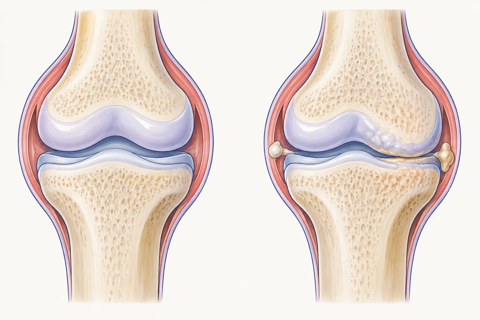
- Uneven cartilage loss
- Joint-space narrowing
- Marginal osteophyte
Smooth articular surfaces
Cartilage covers the bone ends and helps the joint move smoothly.
Osteoarthritis changes
Cartilage can become thinner and uneven, and small bony outgrowths called osteophytes can form.
Remember: Structural changes do not tell the whole story of pain or function. Explain the value of tailored strengthening and aerobic activity as part of the management plan.
Simplified illustration, not to scale. Sources: NHS: osteoarthritis · NICE NG226: osteoarthritis management
Diagnosis (NICE Criteria)
Diagnose clinically if a person:
- • Is aged 45 or over
- • Has activity-related joint pain
- • Has either no morning joint-related stiffness or stiffness that lasts no longer than 30 minutes.
Note: X-rays are not routinely needed and do not correlate well with symptoms.
Management
- Core Treatments (For all): Holistic assessment, education, weight loss (if BMI >25), muscle strengthening and aerobic exercise.
- Pharmacological: Topical NSAIDs (for knee/hand OA) → Oral NSAIDs (co-prescribe PPI) → Opioids (avoid strong opioids if possible). Intra-articular steroid injections can provide short-term relief.
- Surgical: Referral for joint replacement when symptoms substantially impact quality of life despite non-surgical management.
4Rheumatoid Arthritis (RA) and Inflammatory Arthritis
Early diagnosis and treatment with Disease Modifying Anti-Rheumatic Drugs (DMARDs) prevent permanent joint destruction.
Clinical Features
Symmetrical polyarthritis (small joints of hands/feet usually), significant morning stiffness (>60 mins), "boggy" swelling of joints. Positive "Squeeze Test" (metacarpophalangeal/metatarsal joints). Systemic upset (fatigue, flu-like).
Investigations
Rheumatoid Factor (RF), Anti-CCP (more specific), ESR/CRP. Note: Negative RF does not exclude RA (Seronegative RA).
Urgent Referral
Suspected persistent synovitis requires referral to Rheumatology (often a specific Early Arthritis Clinic) ideally within 3 working days of presentation.
Primary Care
Do not delay referral for blood results. Provide analgesia/NSAIDs. Avoid starting oral steroids before specialist assessment as this masks signs.
DMARD Monitoring
GPs often manage repeat prescribing/monitoring (bloods) for Methotrexate, Sulfasalazine, etc., under shared care protocols. Be alert for neutropenia, liver toxicity, and pneumonitis (breathlessness).
5Gout and Crystal Arthropathies
Caused by urate crystal deposition. Often affects the First MTP joint (Podagra).
Acute Attack
Severe pain, redness, swelling (maximal intensity within 12-24 hours).
Treatment: High-dose NSAID (e.g., Naproxen), Colchicine (500mcg bd/tds), or Oral Steroids (Prednisolone 30-35mg x 5 days).
Avoid
Do not start Allopurinol during an acute attack (but continue if already taking).
Prophylaxis (Urate Lowering Therapy - ULT)
- Indications: >1 attack in 12 months, tophi, renal stones, or chronic kidney disease.
- First-line: Allopurinol. Start low (100mg) and titrate up every 4 weeks until Serum Urate target is reached (Target is <360 µmol/L).
- Cover: Co-prescribe Colchicine or NSAID for up to 6 months when starting Allopurinol to prevent flare-ups.
6Polymyalgia Rheumatica (PMR)
Inflammatory condition affecting older adults (>50).
Clinical Features
Bilateral pain and stiffness in the shoulder and/or pelvic girdles. Morning stiffness >45 minutes. Systemic symptoms (fatigue, weight loss).
Investigations
ESR and CRP are almost universally raised. Normal CK (excludes myositis).
Red Flags
Always screen for Giant Cell Arteritis (GCA) (headache, jaw claudication, visual changes).
Management
- Steroids: Dramatic response to Prednisolone 15mg daily (usually within 1 week) confirms diagnosis.
- Tapering: Slow reduction over 1-2 years (e.g., reduce by 2.5mg every 4-8 weeks).
- Bone Protection: Prescribe bisphosphonate + Calcium/Vit D for all patients starting long-term steroids (if age >65 or history of fractures).
7Osteoporosis and Fragility Fractures
A "silent disease" often diagnosed after a low-trauma fracture (fall from standing height or less).
Risk Assessment
Use FRAX or QFracture tools for all women >65, men >75, or younger patients with risk factors (steroids, anorexia, malabsorption, family history, early menopause).
Diagnosis
DEXA scan. T-score ≤ -2.5 = Osteoporosis. T-score between -1.0 and -2.5 = Osteopenia.
Management
- Lifestyle: Weight-bearing exercise, stop smoking, reduce alcohol, adequate Calcium/Vit D intake.
- Pharmacology: Oral Bisphosphonates (Alendronic Acid 70mg weekly) usually first-line. Take on empty stomach, upright for 30 mins.
- Side Effects: GI upset/reflux. Rare: Osteonecrosis of the jaw (dental check needed before starting), Atypical femoral fractures.
8Shoulder Pain: Frozen Shoulder and Rotator Cuff Disorders
Differentiating based on range of movement (ROM).
Frozen Shoulder (Adhesive Capsulitis)
- Features: Pain followed by significant restriction of both active and passive movement (especially external rotation). Associated with Diabetes.
- Phases: Freezing (painful) → Frozen (stiff) → Thawing (recovery). Can take 18-24 months.
- Management: Analgesia, physiotherapy, intra-articular steroid injections (early stage), distension arthrogram.
Rotator Cuff Disorders (Impingement/Tears)
- Features: Pain on overhead activity, painful arc (60-120 degrees). Active movement restricted/painful, but passive movement usually full (unlike frozen shoulder).
- Management: Rest, NSAIDs, Physio (first-line). Steroid injections. Surgery for full-thickness tears (acute traumatic tears in young active patients require urgent referral).

9Hand and Wrist Conditions
Common specific presentations in primary care.
Carpal Tunnel Syndrome
Compression of median nerve. Pain/tingling in thumb/index/middle finger. Worse at night (waking to "shake hand out"). Thenar wasting is a late sign.
Treatment: Wrist splints (night), steroid injection, surgical decompression.
Trigger Finger
Locking/clicking of finger/thumb on flexion. Tendon nodule may be palpable.
Treatment: Avoidance, steroid injection (often curative), surgery.
De Quervain's Tenosynovitis
Pain at the base of the thumb/radial styloid. Positive Finkelstein's test (pain on ulnar deviation with thumb held in fist). Common in new mothers (lifting baby).
Treatment: Splinting, steroid injection.
10Chronic Pain and Fibromyalgia
Defined as pain persisting beyond normal tissue healing time (>3 months).
Visual explanation
One possible cycle in persistent pain
1
Pain with movement
Activity feels uncomfortable, and the person may be unsure what is safe.
2
Worry about harm
Pain may be interpreted as a sign that movement will cause more damage.
3
Less activity
The person avoids movement or withdraws from activities that matter to them.
4
Less confidence and function
Everyday tasks can become harder, reinforcing concern about movement.
Harder tasks can reinforce worry and further avoidance of activity.
Remember: Pain is real and has many contributors. Explore the person’s concerns without blame. Following assessment, agree manageable activity goals and review function and confidence together.
Simplified teaching diagram. Sources: NHS: ways to reduce persistent pain · NICE NG193: assessing and managing chronic pain
Fibromyalgia
Chronic widespread pain, fatigue, unrefreshing sleep, and cognitive symptoms ("fibro-fog"). Diagnosis of exclusion.
Approach: Validate the patient's pain. Shift focus from "cure" to "improving function/quality of life".
Non-Pharmacological
Graded exercise therapy, CBT, sleep hygiene.
Pharmacological
Amitriptyline, Duloxetine, Pregabalin (be cautious of dependency). Opioids are not recommended for chronic primary pain (NICE NG193) as harms often outweigh benefits.
Practice with AI Actors
5 relevant roleplay cases
Apply what you've learned about Musculoskeletal in a realistic, voice-driven consultation. Speak with the AI Actor as you would a real patient and get instant feedback.


Related Topics
Cardiology
Heart disease, hypertension, chest pain, arrhythmias, heart failure and cardiovascular risk assessment.
Respiratory
Asthma, COPD, pneumonia, breathlessness, cough and respiratory infections in primary care.
Gastroenterology
Abdominal pain, IBS, IBD, dyspepsia, liver disease and gastrointestinal red flags.